Effects of Nanotextured PDMS on Cell Culture and Growth

ty10086 提交于 周四, 08/26/2021 - 13:03
文章英文标题
Effects of Nanotextured PDMS on Cell Culture and Growth
正文
Cell adhesion, morphology and growth are known to be influenced by microscale topography of the surfaces. Taking this a step further, nanotextured polydimethylsiloxane (PDMS) substrates were fabricated by micro reactive ion etching (Micro-RIE).This simple process gave good control on surface texture to improve the affinity of PDMS to enhance cell growth. The effects of various nanoscale roughnesses on cell growth were also quantified. Three different recipes were used to prepare nanotextured PDMS by micro-RIE using oxygen (O\u003csub\u003e2\u003c/sub\u003e) and carbon tetrafluoride (CF\u003csub\u003e4\u003c/sub\u003e). First of all etching was performed for 12 min and flow rate of O\u003csub\u003e2 \u003c/sub\u003eand CF\u003csub\u003e4 \u003c/sub\u003ewas kept at a ratio of 1:1 to fabricate \u003citalic\u003esurface 1\u003c/italic\u003e. \u003citalic\u003eSurface 2 \u003c/italic\u003ewas fabricated just by changing the ratio of O\u003csub\u003e2 \u003c/sub\u003eand CF\u003csub\u003e4 \u003c/sub\u003eto 1:2. Then the flow rates of gases were kept same as recipe 1 but the etching time was increased to 25 min to prepare \u003citalic\u003esurface 3\u003c/italic\u003e. Next, surface topography of these PDMS substrates was evaluated quantitatively by measuring root mean square surface roughness using an atomic force microscope (AFM). The measured average roughness of three nanotextured PDMS surfaces were found 242.92±87.709 nm, 515±102.28 nm and 629.17±109.67 nm for \u003citalic\u003esurface 1\u003c/italic\u003e, \u003citalic\u003esurface 2\u003c/italic\u003e and \u003citalic\u003esurface 3\u003c/italic\u003e respectively. The roughnesses of three types of nanotextured surfaces showed a correlation on cell growth. The cell growth rate on flat PDMS surfaces is known to be very low but nanotextured PDMS surfaces allowed exciting cell growth for three different types of cells (human glioblastoma (hGBM), astrocytesand fibroblast cells).The cells were seeded on various surfaces coated with laminin and incubated for 3 days at 37\u003csup\u003eo\u003c/sup\u003eC. The cell densities of experimental surfaces were calculated from their images using \u003citalic\u003eImageJ \u003c/italic\u003esoftware. For all types of cells, growth was higher on nantoextured PDMS surfaces compare to flat PDMS surface and \u003citalic\u003esurface 3\u003c/italic\u003e exhibited highest cell density among three nanotextured surfaces. However density of astrocytes cells was 114 ± 16.15 cells per mm\u003csup\u003e2\u003c/sup\u003e in \u003citalic\u003esurface 3\u003c/italic\u003e where as flat PDMS had only 28 ± 3.58 cells per mm\u003csup\u003e2\u003c/sup\u003e. But standard well plate had higher cell density compare to \u003citalic\u003esurface 3\u003c/italic\u003e and it was 140 ± 18.07 cells per mm\u003csup\u003e2\u003c/sup\u003e. Again density of fibroblasts cells in \u003citalic\u003esurface 3\u003c/italic\u003e was 272.80 ± 13.68 cells per mm\u003csup\u003e2 \u003c/sup\u003ewhich was higher compare to flat PDMS but lower than standard well plate. On the other hand \u003citalic\u003esurface 3\u003c/italic\u003e exhibited very similar cell density as standard well plate for hGBM cells and cell densities were 346 ± 70.64 cells per mm\u003csup\u003e2\u003c/sup\u003e and 334.67 ± 63.23 cells per mm\u003csup\u003e2\u003c/sup\u003e respectively. Consequently it can be deduced that cell growth strongly depends on surface nanotexturing. By comparing three nanotextured surfaces it was found that \u003citalic\u003esurface 1\u003c/italic\u003e exhibited lowest roughness compare to \u003citalic\u003esurface 2\u003c/italic\u003e and \u003citalic\u003esurface 3\u003c/italic\u003e. Therefore available surface area for laminin attachment was lowest in this surface. But \u003citalic\u003esurface 2\u003c/italic\u003e and \u003citalic\u003esurface 3\u003c/italic\u003e had higher roughness and therefore provided larger surface area to attach more laminin. As cell growth is a direct function to available laminin attached in the surface therefore cell density was more in later surfaces. In addition to that circulating tumor cells (CTCs) are known to have very high affinity towards nanotextured surfaces. Therefore density of only hGBM cells in nanotextured surface was very similar to cell density in standard well plate. As a result nanotextured PDMS may be thus useful for cancer cytology devices, especially to culture isolated CTCs for further investigation in microfluidic platforms.;[Formula]
文章内容(英文)
Cell adhesion, morphology and growth are known to be influenced by microscale topography of the surfaces. Taking this a step further, nanotextured polydimethylsiloxane (PDMS) substrates were fabricated by micro reactive ion etching (Micro-RIE).This simple process gave good control on surface texture to improve the affinity of PDMS to enhance cell growth. The effects of various nanoscale roughnesses on cell growth were also quantified. Three different recipes were used to prepare nanotextured PDMS by micro-RIE using oxygen (O\u003csub\u003e2\u003c/sub\u003e) and carbon tetrafluoride (CF\u003csub\u003e4\u003c/sub\u003e). First of all etching was performed for 12 min and flow rate of O\u003csub\u003e2 \u003c/sub\u003eand CF\u003csub\u003e4 \u003c/sub\u003ewas kept at a ratio of 1:1 to fabricate \u003citalic\u003esurface 1\u003c/italic\u003e. \u003citalic\u003eSurface 2 \u003c/italic\u003ewas fabricated just by changing the ratio of O\u003csub\u003e2 \u003c/sub\u003eand CF\u003csub\u003e4 \u003c/sub\u003eto 1:2. Then the flow rates of gases were kept same as recipe 1 but the etching time was increased to 25 min to prepare \u003citalic\u003esurface 3\u003c/italic\u003e. Next, surface topography of these PDMS substrates was evaluated quantitatively by measuring root mean square surface roughness using an atomic force microscope (AFM). The measured average roughness of three nanotextured PDMS surfaces were found 242.92±87.709 nm, 515±102.28 nm and 629.17±109.67 nm for \u003citalic\u003esurface 1\u003c/italic\u003e, \u003citalic\u003esurface 2\u003c/italic\u003e and \u003citalic\u003esurface 3\u003c/italic\u003e respectively. The roughnesses of three types of nanotextured surfaces showed a correlation on cell growth. The cell growth rate on flat PDMS surfaces is known to be very low but nanotextured PDMS surfaces allowed exciting cell growth for three different types of cells (human glioblastoma (hGBM), astrocytesand fibroblast cells).The cells were seeded on various surfaces coated with laminin and incubated for 3 days at 37\u003csup\u003eo\u003c/sup\u003eC. The cell densities of experimental surfaces were calculated from their images using \u003citalic\u003eImageJ \u003c/italic\u003esoftware. For all types of cells, growth was higher on nantoextured PDMS surfaces compare to flat PDMS surface and \u003citalic\u003esurface 3\u003c/italic\u003e exhibited highest cell density among three nanotextured surfaces. However density of astrocytes cells was 114 ± 16.15 cells per mm\u003csup\u003e2\u003c/sup\u003e in \u003citalic\u003esurface 3\u003c/italic\u003e where as flat PDMS had only 28 ± 3.58 cells per mm\u003csup\u003e2\u003c/sup\u003e. But standard well plate had higher cell density compare to \u003citalic\u003esurface 3\u003c/italic\u003e and it was 140 ± 18.07 cells per mm\u003csup\u003e2\u003c/sup\u003e. Again density of fibroblasts cells in \u003citalic\u003esurface 3\u003c/italic\u003e was 272.80 ± 13.68 cells per mm\u003csup\u003e2 \u003c/sup\u003ewhich was higher compare to flat PDMS but lower than standard well plate. On the other hand \u003citalic\u003esurface 3\u003c/italic\u003e exhibited very similar cell density as standard well plate for hGBM cells and cell densities were 346 ± 70.64 cells per mm\u003csup\u003e2\u003c/sup\u003e and 334.67 ± 63.23 cells per mm\u003csup\u003e2\u003c/sup\u003e respectively. Consequently it can be deduced that cell growth strongly depends on surface nanotexturing. By comparing three nanotextured surfaces it was found that \u003citalic\u003esurface 1\u003c/italic\u003e exhibited lowest roughness compare to \u003citalic\u003esurface 2\u003c/italic\u003e and \u003citalic\u003esurface 3\u003c/italic\u003e. Therefore available surface area for laminin attachment was lowest in this surface. But \u003citalic\u003esurface 2\u003c/italic\u003e and \u003citalic\u003esurface 3\u003c/italic\u003e had higher roughness and therefore provided larger surface area to attach more laminin. As cell growth is a direct function to available laminin attached in the surface therefore cell density was more in later surfaces. In addition to that circulating tumor cells (CTCs) are known to have very high affinity towards nanotextured surfaces. Therefore density of only hGBM cells in nanotextured surface was very similar to cell density in standard well plate. As a result nanotextured PDMS may be thus useful for cancer cytology devices, especially to culture isolated CTCs for further investigation in microfluidic platforms.;[Formula]
来源出处
Journal|[J]Meeting AbstractsVolume MA2014-01, Issue 43. 2014. PP 1611-1611

PDMS臻品推荐

PDMS-微流控基质材料-PDMS/道康宁SYLGARD184-聚二甲基硅氧烷/0.5KG(组)

产品说明书PDF自助在线看:http://www.pdmshub.com/sih

信息更新:

美国总工厂生产的0.5KG原始包装的PDMS延期至2021年9月16日后才能交货,默认我们将发国内库存的进口分装产品(02085925-0.5KG);另受制于有机硅大幅涨价潮影响,2021年9月1日后将上调销售价格,另行通知。Dow Corning=道康宁=Dow SiL=陶氏,都是同一家公司。 2021.09.01更新

美国总工厂生产的0.5KG包装的PDMS恢复正常进口,合法正规,质量金标准,欢迎订购使用。并承诺从我司购买的皆为原厂包装,100%未开封,附带技术手册,非国产灌装仿制、非分装、非仿冒外包装产品,可以放心使用。大量订购更有优惠。2021.04.01更新

备注:由于dowsil与道康宁公司合并,美国总工厂生产的PDMS,价格涨幅高达1倍之多,因针对科研用户使用,特备货规格0.5KG。如果您需要美国总工厂原厂生产的大规格包装的预定,可以与QQ 11366508联系给予报价/货期。2021.05更新

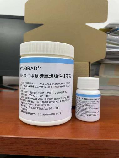
此链接为PDMS-微流控芯片/PDMS/道康宁SYLGARD184/小桶0.5KG装的链接。

一般性的产品性能参数表参考以下小桶装的参数:

微流控芯片/PDMS/道康宁SYLGARD184;微流控芯片/PDMS/道康宁SYLGARD184;微流控芯片/PDMS/道康宁SYLGARD184;

品牌型号:道康宁SYLGARD184

包装规格:0.5KG/罐[含有45.4g固化剂,总重量为0.5KG]

产品颜色:保质期限:36个月

存放环境说明:室温,阴凉处保存

备注说明:

美国道康宁道康宁SYLGARD 184硅橡胶是由液体组分组成的双组分套件产品,包括基本组分与固化剂。基本组分与固化剂按10:1重量比完全混合,中等粘度混合液的稠度与SAE 40机油相似。无论厚薄,混合液将固化成为具有韧性的透明弹性体,最适用于电子/电气方面的封装与灌封应用。 道康宁SYLGARD 184硅橡胶在25~150℃的温度范围内固化,无放热现象,无需二次固化。固化过程完成后,可立即在-55~200℃的温度范围内使用。 产品特性:低毒性,在常规的工业操作中,无特别的注意事项; 无溶剂或固化副产物, 固化时不放热;无需特殊的通风条件,不会产生腐蚀;固化时,收缩量小; 固化后, 透明具有弹性;抗震与减缓机械震动;振动的传递性能小;元件可裸视检查与易修补性; 环保性能;低吸水性,良好的耐辐射性能;高真空状态下的低漏气性; 优异的电性能;较大温度范围内的稳定性, 抗解聚;在-55~200℃范围内,甚至在密闭状态下保持弹性与柔韧性,性能稳定; 阻燃性,UL可燃性分级为94 V-1,温度等级:130℃ 产品用途: 道康宁SYLGARD 184硅橡胶在电气/电子的封装与灌封方面有广泛的应用。